Tag Archives: neurology

WHYCAN’TIHITYOU?!


Currently, there is a housefly buzzing around my head. Every single time it lands I attempt this futile clapping motion to destroy it. I fail. I fail time and time again. So, my question is, obviously, WHY CAN’T I HIT YOU!?

After a small amount of Google-sleuthing I found the answer here. It seems Drosophila contain a pair of large aptly named nerves, Giant Fiber. This bundle runs the entire length of the head and down to the thorax. At the endpoint it triggers the thoracic ganglion which then shoots elsewhere. What triggers this giant bundle to begin with? The eyes! It uses visual cues to initiate its escape sequence. I wonder why it would be associated with something like that…

Just an eye... NBDSo if the Giant Fibers end with the thoracic ganglion what happens next? The ganglion shoots the signal to the dorsal longitudinal muscle (DLM) and tergotrochanteral muscle (TTM or “jump muscles”). This moves two thing: the legs and the wings. What do the legs do? JUMP! Thus, the reason they’re called jump muscles. The wings do something a little more complex. Upon receiving a signal  the wings go from the closed position to the open position and slightly elevate. So really the one nerve bundle initiates a double whammy of legs and wings. The strange part is that the TTM does both of these functions. The DLM is only indirectly involved.

So the take away message is that a simple little pathway is why I can’t kill this damn fly.

Sidenote, the paper is a little dated as it was published in 1983 but the general workings are still the same.

*Update*
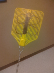
Flies didn’t evolve around flyswatters. Gotcha.Owned.


Notch and Delta in a Nutshell


Cell signaling is a very important process within multi-cellular organisms. Yet, many people grossly misunderstand how cells could communicate with one another. During the Answers in Genesis conference that stopped by Morris last year, one individual was under the impression that “cellular e-mail” must exist. “Why? Well, because it must!” *sigh* So, the pathway that I’m going to look at is Notch. Why? Well, because I must!  It’s also pretty cool, I swear.

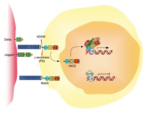
So here we have the Notch pathway. http://www.humpath.com/IMG/jpg_notch_jagged_gs_01.jpg Notch is a transmembrane protein which means it goes straight through the cell membrane and protrudes on either side. This protein acts as a form of hair-trigger. When activated it cleaves the inside part which heads off to the nucleus to play with gene expression. Notch isn’t activated by just any protein flying into it, and this is where Delta comes into play. Now the specifics vary depending on species (humans don’t actually have Delta but it’s similar and therefore Delta-like) but the purpose remains relatively the same.

**Now, I’m grossly oversimplifying the Notch pathway. I do this not because the areas I’m overlooking are not important (they are!), but this is a neurology post and it’s far too easy to get bogged down in minutia of everything and overlook the cool stuff of one area. **

So Delta and Notch touch, part of Notch breaks off and heads off into the cell to make shenanigans. What does this shenaniganary have to do with cell signaling and the nervous system? These have two functions. One is to adhere the two cells to one another. The other function is in development, in this case neuro-development. Once the cell with Delta contacts the Notch cell the piece inside breaks off and heads for the nucleus. What it does here is stop the cell from differentiating while the Delta cell remains able to do so. Notch inhibits rather than induces. It’s a bit backwards from what you would expect, but the role of Notch in neural development is to say who cannot become neural tissue.  As for the Delta cell, eventually it will move out of the epithelial layer and differentiate into a neuroblast.

If you think about it, this makes sense. If something induces neural differentiation then something should also stop it to avoid something made entirely of nervous system. Now, I can’t emphasize enough that Notch does other things. Those things just reach beyond the scope of this class. This is just a quick look at how two little things poking out of a cell can have a big impact.


The Role of Neuroplasticity in Constraint-Induced Therapy


WOOHOO! I am finally done with my senior paper. As you may surmise from the title I chose to write it on the neuro-mechanics involved with CI therapy. It’s written with a more informal tone but nonetheless it still landed me high marks.  Comments, questions, and retweets (#revfrost) are always welcome. Enjoy!

ABSTRACT: The human brain is capable of extraordinary change. The impact of strokes can be devastating and often times seen as refractory. Constraint Induced (CI) therapy has proved beneficial in alleviating common symptoms caused by strokes. Recent evidence provides an insight in identifying the biological mechanisms exploited by CI-therapy: neurogenesis, gliosis, and synaptogenesis.

Continue reading


A Brief History of the Brain: Ancient Egyptians


The brain has always been looked upon with reverence and awe for its power and capacity of conscious thought… right? Absolutely not. Not only is the brain the coolest thing in science (no bias, I swear) but historically it has been neglected or set aside as the “housing of the soul.” Well, that’s irritating.

So let’s start back in ye’ olden days of the pharaohs. It seems the Egyptians fall into a weird category of belief. They recognized the problems that would arise from head wounds but ignored it in terms of thought and consciousness. During the mummification process they would enter the nasal cavity, break through the thin-layered ethmoid bone, and draw out the contents of the skull. We’ve all heard this spiel since elementary school. The important parts to ask though are the why and how.

The why seems relatively straight forward. It was removed along with all the other organs excluding the heart to prepare the dead for their afterlife. The heart was left in place to be judged in Duat, the Egyptian underworld. Under supervision of Anubis, the heart was weighed against Ma’at’s feather of truth. The Declaration of Innocence: Spell 125 details a 42-part moral system which they were judged upon – similar to that of the 10 Commandments of Christianity but 2000 years prior (coincidence? >_>). If the heart was found to be unworthy it was eaten by the “devourer”, Ammut. If not, they proceeded to Aaru, the heavenly reed field.

That’s neat and all, but it’s not the whole story. Only 50% of recovered mummy skulls had the brain removed prior to mummification. In addition, the eyes had also been removed and replaced with various materials. Why? Because they rot and stay moist. The whole point of mummification is to remove as much water as possible to preserve the remaining tissue. The body needed to stay as recognizable as possible; I’ll explain in a minute. An example concoction used as a dehydrant consisted of 84.7% sodium carbonate or bicarbonate, 1.5% sodium chloride, and 13.8% sodium sulphate. While this prevents most bacterial decay it doesn’t stop it all. The brain and eyes start to leak fluid and rot. Quick fix? Take them out. Sure, it’s a little more difficult than the other organs, but it works better in the long-run. A dry mummy is a happy mummy.Credit goes to http://www.offbeatenough.com/

Another key part is how the brain was removed. Mummification was usually performed in a manner that minimized cosmetic damage as it was believe the soul (the Ba) needed to recognize the body to return to it every night. The  common belief of the process behind it seems to be a gross misconception. It is largely held that the brain was sucked out, scooped out with a hook or some variation thereof. For the most part, this is untrue. The tool used did indeed have a hook but it did not function in that fashion. The viscosity of the brain allowed it to stick to the entire tool. So, as the tool would be drawn out it would bring brain with it. With repeated entry from the tool the inside tissue would begin to liquefy and eventually could be poured out of the nasal cavity.

The Egyptians were pioneers of the medical realm. They were even recognized by Homer in the Odyssey: “In Egypt, the men are more skilled in medicine than any of human kind.” The Edwin Smith Surgical Papyrus details a great many of their accomplishments along with a few of their more… unique practices. Maybe with a bit more time they could have perfected the art of medicine a little more. Until next time, remember to “pour milk into both ears” for a wounded temple.

The Problem of Brain Removal during Embalming by the Ancient Egyptians
F. Filce Leek. The Journal of Egyptian Archaeology

Dynamic Neuroplasticity after Human Prefrontal Cortex Damage.


ResearchBlogging.org In this article, the authors focused on the effects of a unilateral lesion within the prefrontal cortex. Human vision works upon a rather backwards system. Information from the left side of each eye moves toward the left occipital lobe to be analyzed. The right follows an identical pattern.

So essentially, two sides of the eyeball swap information between hemispheres. From here, the information is sent forward along the current hemisphere to be analyzed by the prefrontal cortex (PFC).Because what else would you expect from the brain? In a nutshell, the left and right fields of vision (of a single eye) are interpreted by different halves of the brain. This is where the problems begin to arise. If the lesion occupies the left PFC, what happens to that information? Does it simply get left unanalyzed and you lose that field in each eye?

In a static environment you could assume that the side affected by the lesion would lose greater processing for that side of the brain. However, if the brain were dynamic and could re-route the circuit, information from that field could have the potential to return. It seems that’s just what occurs but with a small cost. The information destined for the left PFC follows the tradition route back to the prefrontal cortex but stops short, makes a pass through the corpus callosum,  and arrives at the contralateral prefrontal cortex (right side). The right prefrontal cortex actually picks up the slack left from the damaged area. However, as this seems almost too good to be true, this is not the case under all conditions. When an object is detected in the affected field compensation occurs. Yet, when objects are present to both fields simultaneously the right PFC solely analyzes the information from the right field. In other words, the right PFC picks up the slack so long as it has no incoming information itself.

The brain undergoes this change after repeated exposure to left field stimuli on a trial-by-trial basis. While the left PFC never fully recovers, this example shows a rather interesting compensation mechanism that can be achieved plastically. Although the paper never actually discussed this, I can’t help but wonder about the underlying cellular changes associated with this feat. Currently my assumption would just be an increase in synapses (synaptogenesis) favoring the re-wiring across the corpus callosum and to the contralateral cortex. But that’s just my guess. If anyone has any thoughts to the contrary I would certainly be willing to entertain them.

Voytek, B., Davis, M., Yago, E., Barceló, F., Vogel, E., & Knight, R. (2010). Dynamic Neuroplasticity after Human Prefrontal Cortex Damage Neuron, 68 (3), 401-408 DOI: 10.1016/j.neuron.2010.09.018 

*UPDATE*: I figured using ResearchBlogger would be more efficient.


Circadian Rhythm and Synapse Activity in Zebrafish


The other day I found an article that seemed all too perfect. Development. Neurology. Zebrafish. For those of you who don’t know, these little fishies are not only PZ’s specialty but are also currently taking over our lab (You will be missed, fishies in tank #3).

In zebrafish, the sleep-wake cycle is regulated by hypocretin neurons (HCRT). These neurons also play a major role in mammals as well and if damaged can lead to narcolepsy. The researchers labeled these neurons in zebrafish with a presynaptic marker synaptophysin (SYP) attached to a fluorescent dye. Now the researchers can look at activity across the span of a day to visually quantify changes in baseline function or artificially produced by various conditions (e.g. sleep deprivation). So,  they irritated fish for several hours after normal sleeping time until the fish were nice and sleep deprived.

The sleep deprived fish showed homeostatic influences on the number of HCRT synapses. Why is this important? The fish showed that upon sleep deprivation more synapse terminals were created  to maintain cellular balance. Yet, simultaneously, the natural circadian rhythm began to decrease the number of synapses from the point of initial sleep deprivation. Homeostatic effects are estimated to be a mere 17% and this result appears only after six hours of deprivation. Essentially, as the day wears on the brain winds up more and more. The circadian rhythm counteracts this by gradually resetting the system back to baseline. A portion of this study showed that as the system tries to reset, sleep deprivation will weakly offset efficacy leading to impairment in memory retainment (and overall cranky fish).

*Professor, I would have gotten that question right but the course workload created homeostatic effects which led to faulty memory retainment!*


Sexual Dimorphism in Drosophila


Last week, Current Biology released an article about structural differences between sexes in Drosophila melanogaster. It may not be about a human brain, but it’s still neurology and at least semi-related to developmental biology this time.

While the “model” organism displays distinct behavioral differences between sexes, the overall anatomy in regards to dimorphism has been essentially neglected. Previously, the only discernible difference was in the olfactory system. Male olfactory systems have a 25 – 60% larger volume than female flies. Coincidently the male pheromone cVA attracts females while repelling males (however no credible correlation can be made).

Ooooo pretty!

Pretty picture! This is one of many from a collage presented in the article and most interesting to myself as it visibly shows the gender difference in brain size. Magenta is larger in females and green is larger in males.

The article proceeds to explain the fine tunings of specific proteins (fruitless, sex lethal, and transformer) and their effect on dimorphism in a manner that is, quite frankly, far beyond my level of comprehension.

Overall, sexual dimorphism does exist in the Drosophila brain and may influence behavior as a result. A key example of this lies in the activation of fruM (the male version of the fruitless protein) in females and the resulting behavior of courting other females. Now that the anatomical groundwork has been laid the researchers are able to pursue behavioral consequences in future research.