Category Archives: Science Posts

WHYCAN’TIHITYOU?!


Currently, there is a housefly buzzing around my head. Every single time it lands I attempt this futile clapping motion to destroy it. I fail. I fail time and time again. So, my question is, obviously, WHY CAN’T I HIT YOU!?

After a small amount of Google-sleuthing I found the answer here. It seems Drosophila contain a pair of large aptly named nerves, Giant Fiber. This bundle runs the entire length of the head and down to the thorax. At the endpoint it triggers the thoracic ganglion which then shoots elsewhere. What triggers this giant bundle to begin with? The eyes! It uses visual cues to initiate its escape sequence. I wonder why it would be associated with something like that…

Just an eye... NBDSo if the Giant Fibers end with the thoracic ganglion what happens next? The ganglion shoots the signal to the dorsal longitudinal muscle (DLM) and tergotrochanteral muscle (TTM or “jump muscles”). This moves two thing: the legs and the wings. What do the legs do? JUMP! Thus, the reason they’re called jump muscles. The wings do something a little more complex. Upon receiving a signal  the wings go from the closed position to the open position and slightly elevate. So really the one nerve bundle initiates a double whammy of legs and wings. The strange part is that the TTM does both of these functions. The DLM is only indirectly involved.

So the take away message is that a simple little pathway is why I can’t kill this damn fly.

Sidenote, the paper is a little dated as it was published in 1983 but the general workings are still the same.

*Update*

Flies didn’t evolve around flyswatters. Gotcha.Owned.


Notch and Delta in a Nutshell


Cell signaling is a very important process within multi-cellular organisms. Yet, many people grossly misunderstand how cells could communicate with one another. During the Answers in Genesis conference that stopped by Morris last year, one individual was under the impression that “cellular e-mail” must exist. “Why? Well, because it must!” *sigh* So, the pathway that I’m going to look at is Notch. Why? Well, because I must!  It’s also pretty cool, I swear.

So here we have the Notch pathway. http://www.humpath.com/IMG/jpg_notch_jagged_gs_01.jpg Notch is a transmembrane protein which means it goes straight through the cell membrane and protrudes on either side. This protein acts as a form of hair-trigger. When activated it cleaves the inside part which heads off to the nucleus to play with gene expression. Notch isn’t activated by just any protein flying into it, and this is where Delta comes into play. Now the specifics vary depending on species (humans don’t actually have Delta but it’s similar and therefore Delta-like) but the purpose remains relatively the same.

**Now, I’m grossly oversimplifying the Notch pathway. I do this not because the areas I’m overlooking are not important (they are!), but this is a neurology post and it’s far too easy to get bogged down in minutia of everything and overlook the cool stuff of one area. **

So Delta and Notch touch, part of Notch breaks off and heads off into the cell to make shenanigans. What does this shenaniganary have to do with cell signaling and the nervous system? These have two functions. One is to adhere the two cells to one another. The other function is in development, in this case neuro-development. Once the cell with Delta contacts the Notch cell the piece inside breaks off and heads for the nucleus. What it does here is stop the cell from differentiating while the Delta cell remains able to do so. Notch inhibits rather than induces. It’s a bit backwards from what you would expect, but the role of Notch in neural development is to say who cannot become neural tissue.  As for the Delta cell, eventually it will move out of the epithelial layer and differentiate into a neuroblast.

If you think about it, this makes sense. If something induces neural differentiation then something should also stop it to avoid something made entirely of nervous system. Now, I can’t emphasize enough that Notch does other things. Those things just reach beyond the scope of this class. This is just a quick look at how two little things poking out of a cell can have a big impact.


The Role of Neuroplasticity in Constraint-Induced Therapy


WOOHOO! I am finally done with my senior paper. As you may surmise from the title I chose to write it on the neuro-mechanics involved with CI therapy. It’s written with a more informal tone but nonetheless it still landed me high marks.  Comments, questions, and retweets (#revfrost) are always welcome. Enjoy!

ABSTRACT: The human brain is capable of extraordinary change. The impact of strokes can be devastating and often times seen as refractory. Constraint Induced (CI) therapy has proved beneficial in alleviating common symptoms caused by strokes. Recent evidence provides an insight in identifying the biological mechanisms exploited by CI-therapy: neurogenesis, gliosis, and synaptogenesis.

Continue reading


Dynamic Neuroplasticity after Human Prefrontal Cortex Damage.


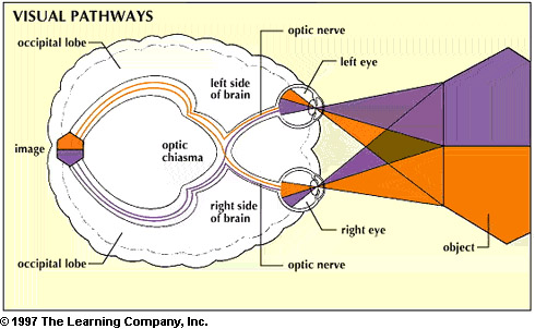
ResearchBlogging.org In this article, the authors focused on the effects of a unilateral lesion within the prefrontal cortex. Human vision works upon a rather backwards system. Information from the left side of each eye moves toward the left occipital lobe to be analyzed. The right follows an identical pattern.

So essentially, two sides of the eyeball swap information between hemispheres. From here, the information is sent forward along the current hemisphere to be analyzed by the prefrontal cortex (PFC).Because what else would you expect from the brain? In a nutshell, the left and right fields of vision (of a single eye) are interpreted by different halves of the brain. This is where the problems begin to arise. If the lesion occupies the left PFC, what happens to that information? Does it simply get left unanalyzed and you lose that field in each eye?

In a static environment you could assume that the side affected by the lesion would lose greater processing for that side of the brain. However, if the brain were dynamic and could re-route the circuit, information from that field could have the potential to return. It seems that’s just what occurs but with a small cost. The information destined for the left PFC follows the tradition route back to the prefrontal cortex but stops short, makes a pass through the corpus callosum,  and arrives at the contralateral prefrontal cortex (right side). The right prefrontal cortex actually picks up the slack left from the damaged area. However, as this seems almost too good to be true, this is not the case under all conditions. When an object is detected in the affected field compensation occurs. Yet, when objects are present to both fields simultaneously the right PFC solely analyzes the information from the right field. In other words, the right PFC picks up the slack so long as it has no incoming information itself.

The brain undergoes this change after repeated exposure to left field stimuli on a trial-by-trial basis. While the left PFC never fully recovers, this example shows a rather interesting compensation mechanism that can be achieved plastically. Although the paper never actually discussed this, I can’t help but wonder about the underlying cellular changes associated with this feat. Currently my assumption would just be an increase in synapses (synaptogenesis) favoring the re-wiring across the corpus callosum and to the contralateral cortex. But that’s just my guess. If anyone has any thoughts to the contrary I would certainly be willing to entertain them.

Voytek, B., Davis, M., Yago, E., Barceló, F., Vogel, E., & Knight, R. (2010). Dynamic Neuroplasticity after Human Prefrontal Cortex Damage Neuron, 68 (3), 401-408 DOI: 10.1016/j.neuron.2010.09.018 

*UPDATE*: I figured using ResearchBlogger would be more efficient.


I Think, Therefore I Am


This was a piece written for our Developmental Biology final. I wrote this with the intention of pushing a radical, controversial concept that I had never seen before. This article does not necessarily reflect my own views. This was merely an assignment I had a bit of evil fun with. Enjoy!

–Rev.Frost

Continue reading


Innate Immunity: So Nice, They Made It Twice


People often use the phrase “do not reinvent the wheel” to describe using an existing method towards a new task rather than creating one anew. Evolution tends to follow a similar philosophy. Many changes utilize genetic toolkits that have long been present, tucked away in the genome of the organism. Sometimes these toolkits have already been used repeatedly for various traits. The common fruit fly (Drosophila melanogaster), a model test subject for over a century, has been scrutinized in remarkable detail to gain an understanding of molecular interactions and evolution. When researchers discovered the NF-kB (nuclear factor kappa-light-chain-enhancer of activated B cells) pathway in mammals that functioned remarkably similar to the Toll pathway in Drosophila it was assumed that the immunological pathways were homologous (e.g. using the same toolkit).

The NF-kB pathway establishes its importance due to its swift reaction time. Most transcription factors are synthesized as needed. Building a protein from scratch then applying it where

Figure 1: Contrasting transcription pathways between Drosophila and Mammals

needed takes a fair amount of time. Sometimes, like during a pathogenic invasion, the cell cannot survive long enough to produce these proteins. In the immune system these proteins can be constructed preemptively, bound, and inhibited in the cytosol for future use. The NF-kB pathway activates these stored proteins by cleaving the inhibiting molecule and thus allowing the transcription factor, NF-kB, to enter the nucleus. The Toll pathway operates, essentially, in the same fashion as NF-kB (Figure 1). However, Drosophila uses completely different proteins to ultimately provoke a similar immune response with Dorsal-related immunity factor (Dif ).

The Toll pathway plays another, yet significant role only found in Drosophila. After the egg is

Figure 2: The Toll-Dorsal Pathway in Drosophila melanogaster

fertilized it lays the foundation for the rest of development, ventral and dorsal (down and up). Upon fertilization, the ligand Spätzle (SPZ) is distributed across the perivitelline membrane (Figure 2a). From here, SPZ binds to the Toll receptors of cells maternally designated to become ventral. The Toll receptors set in motion a cascade that phosphorylates CACT (the inhibitor) which allows the transcription factor to promote the production of the protein Dorsal (Figure 2b).

These pathways were initially thought to have arisen from a common ancestor to both Drosophila and mammals. However, it seems this is only partially correct. Genomic research opened up new, previously unexplored areas to consider. This pathway did, initially, form in a Eumetazoan ancestor. This pathway was markedly underdeveloped and only contained a few components. When the bilateral lineage diverged into deuterostomes and protostomes the common pathway ended. From here, both NF-kB and Toll evolved independently through gene duplication.

These diverse differences between pathways give great insight to the evolutionary history of the circuit itself. In order to have diverged so far in composition yet retain similar function suggests the ancestral pathway to have been particularly modular. In other words, the ancestral pathway was constructed using multiple pieces (modules). This setup allows individual modules to be modified over time without removing the core process of the system. Drosophila slowly modified this pathway to affect larval development by introducing Dorsal and SPZ into the circuit. As a result, Drosophila is able to use a single genetic circuit for dual regulation.

In Biology, nothing is ever as simple as it appears to be. The subjects could range from immune response pathways and developmental regulation to simple autosomal point mutations, but more research is always needed. The NF-kB pathway was thought to be well known until just recently, yet new information radically altered our understanding of it. With additional research, who knows what we could uncover next?

Belvin, M. P., Anderson, K. V., 1996. A CONSERVED SIGNALING PATHWAY: The Drosophila Toll-Dorsal Pathway. Annual Review Cell Developmental Biology. 12, 396-416.

Lemaitre, B., 2004. The road to Toll. Nature Reviews Immunology. 4, 521-527.

Leulier, F., Lemaitre, B., 2008. Toll-like receptors — taking an evolutionary approach. Nature Reviews Genetics. 9, 165-178.

Waterhouse, R. M., Kriventseva, E. V., Meister, S., Xi, Z., Alvarez, K. S., Bartholomay, L. C., Barillas-Mury, C., Bian, G., Blandin, S., Christensen, B. M., Dong, Y., Jiang, H., Kanost, M. R., Koutsos, A. C., Levashina, E. A., Li, J., Ligoxygakis, P., MacCallum, R. M., Mayhew, G. F., Mendes, A., Michel, K., Osta, M. A., Paskewitz, S., Shin, S. W., Vlachou, D., Wang, L., Wei, W., Zheng, L., Zou, Z., Severson, D. W., Raikhel, A. S., Kafatos, F. C., Dimopoulos, G., Zdobnov, E. M., Christophides, G. K., 2007. Evolutionary Dynamics of Immune-Related Genes and Pathways in Disease-Vector Mosquitoes. Science. 316, 1738-1743.

Trisomy…22?


Many people are familiar with Trisomy 21 in humans. Commonly known as Down Syndrome, Trisomy 21 severely affects human development via complex gene and environmental interactions. Genetically speaking, is this condition novel to humans? Or can this be seen in other primates? Most of the “great apes” contain 24 pairs of chromosomes, humans being the exception with 23. When compared, the two karyotypes  look strikingly similar. The only vast difference occurring on chromosome 2. From the diagram below, the human chromosome 21 and the great ape chromosome 22 look remarkably similar. Documented cases of Trisomy 22 in chimpanzees have been seen as far back as 1969. With the additional #22, symptoms parallel to Down Syndrome appeared. Continue reading


Circadian Rhythm and Synapse Activity in Zebrafish


The other day I found an article that seemed all too perfect. Development. Neurology. Zebrafish. For those of you who don’t know, these little fishies are not only PZ’s specialty but are also currently taking over our lab (You will be missed, fishies in tank #3).

In zebrafish, the sleep-wake cycle is regulated by hypocretin neurons (HCRT). These neurons also play a major role in mammals as well and if damaged can lead to narcolepsy. The researchers labeled these neurons in zebrafish with a presynaptic marker synaptophysin (SYP) attached to a fluorescent dye. Now the researchers can look at activity across the span of a day to visually quantify changes in baseline function or artificially produced by various conditions (e.g. sleep deprivation). So,  they irritated fish for several hours after normal sleeping time until the fish were nice and sleep deprived.

The sleep deprived fish showed homeostatic influences on the number of HCRT synapses. Why is this important? The fish showed that upon sleep deprivation more synapse terminals were created  to maintain cellular balance. Yet, simultaneously, the natural circadian rhythm began to decrease the number of synapses from the point of initial sleep deprivation. Homeostatic effects are estimated to be a mere 17% and this result appears only after six hours of deprivation. Essentially, as the day wears on the brain winds up more and more. The circadian rhythm counteracts this by gradually resetting the system back to baseline. A portion of this study showed that as the system tries to reset, sleep deprivation will weakly offset efficacy leading to impairment in memory retainment (and overall cranky fish).

*Professor, I would have gotten that question right but the course workload created homeostatic effects which led to faulty memory retainment!*


Sexual Dimorphism in Drosophila


Last week, Current Biology released an article about structural differences between sexes in Drosophila melanogaster. It may not be about a human brain, but it’s still neurology and at least semi-related to developmental biology this time.

While the “model” organism displays distinct behavioral differences between sexes, the overall anatomy in regards to dimorphism has been essentially neglected. Previously, the only discernible difference was in the olfactory system. Male olfactory systems have a 25 – 60% larger volume than female flies. Coincidently the male pheromone cVA attracts females while repelling males (however no credible correlation can be made).

Ooooo pretty!

Pretty picture! This is one of many from a collage presented in the article and most interesting to myself as it visibly shows the gender difference in brain size. Magenta is larger in females and green is larger in males.

The article proceeds to explain the fine tunings of specific proteins (fruitless, sex lethal, and transformer) and their effect on dimorphism in a manner that is, quite frankly, far beyond my level of comprehension.

Overall, sexual dimorphism does exist in the Drosophila brain and may influence behavior as a result. A key example of this lies in the activation of fruM (the male version of the fruitless protein) in females and the resulting behavior of courting other females. Now that the anatomical groundwork has been laid the researchers are able to pursue behavioral consequences in future research.


Cerebral Palsy


Last week I received a request from MarkNS asking about cerebral palsy therapy when viewed from neuroplasticity. Sounds good, let’s look at that.

I did not know much about cerebral palsy prior to this post, but strangely I do know an ultimate fighter with it…  Cerebral palsy is a multi-faceted disorder that comes in all shapes in sizes, many of which are outside the scope of this post. When looked at with a simplistic view, cerebral palsy is a movement disorder caused by damage to the motor cortex of the brain.

Brain damage. “Irreversible. Permanent.” <–Wrong! The most common form of brain damage associated with neuroplasticity is in stroke patients. This is where the greater body of research can be found. It would seem that for many years all signs pointed to “you’re screwed”.  That is, until Edward Taub came onto the scene.

Taub, through deafferentation experiments on monkeys, invented a new form of therapy, Constraint Induced (CI) Therapy (Simple or Advanced explanation, I recommend the advanced if you have the time as I will only touch lightly on it). The premise of this therapy focuses on a phenomenon called “learned non-use”. In essence, after losing a degree of control in one limb you then compensate with the other to pick up the slack. (e.g. after a stroke affecting the left side, you would use your right side that much more.) When this compensation occurs the neural pathways begin to weaken, leading to even further non-use.

So we know how to lose it, now how do we get it back? CI Therapy essentially forces the affected limb to switch roles. By restraining the “good” limb and using only the “bad” one,  the patient begins to build motor control on the weaker side. It turns out this not only builds character but rebuilds the pathways!

So that’s all fine and dandy for strokes, but what about cerebral palsy? There’s an app for that. Unfortunately the only data I could find was done on children with hemiparesis associated with cerebral palsy. The good news is that the children responded amazingly well to the therapy and sustained the results afterwards. The range of effectiveness is still limited for the moment. CI Therapy has come a long way and it would be a safe assumption that it will continue on.