Monthly Archives: September 2011

A Continuous-Time Neural Network Model


Here’s a paper, co-authored by one of my frequently read neurologists Merzenich, regarding the brain’s ability to display sensory input in sync with time. Previously, studies only examined tasks that utilized spatially differentiated patterns. These studies neglected the influence of excitatory and inhibitory postsynaptic potential summation (ePSPs/iPSPs). Why did the authors choose these neural properties? It was a shot in the dark essentially. While well characterized, no one really knows how these contribute to information processing. This seems like a good way to find out.

They proceeded with a few sets of experiments. They examined ePSPs set with paired-pulse facilitation (PPF), iPSPs with PPF, and PPF with slow iPSPs. In the last category, they saw that when they sent two identical sequential signals down the same input channel the second reaction varied from the first. In fact, they saw that 25-50% of one cell layer showed time-sensitive behavior.

From here the authors fine tune their data using multiple time intervals as well as varying frequencies of the stimulus. The take home message here is that no major reconstruction of previous models was needed. All that was needed was to observe these overlooked properties in the proper context. The authors hope that with increasing complexity of study and incorporating a few other properties, like Hebbian plasticity, will further improve our understanding the transformation of temporal signaling into spatial signaling.

Perhaps next post I’ll take a look at Hebbian plasticity to better understand where this line of research could go. Cheers.


WHYCAN’TIHITYOU?!


Currently, there is a housefly buzzing around my head. Every single time it lands I attempt this futile clapping motion to destroy it. I fail. I fail time and time again. So, my question is, obviously, WHY CAN’T I HIT YOU!?

After a small amount of Google-sleuthing I found the answer here. It seems Drosophila contain a pair of large aptly named nerves, Giant Fiber. This bundle runs the entire length of the head and down to the thorax. At the endpoint it triggers the thoracic ganglion which then shoots elsewhere. What triggers this giant bundle to begin with? The eyes! It uses visual cues to initiate its escape sequence. I wonder why it would be associated with something like that…

Just an eye... NBDSo if the Giant Fibers end with the thoracic ganglion what happens next? The ganglion shoots the signal to the dorsal longitudinal muscle (DLM) and tergotrochanteral muscle (TTM or “jump muscles”). This moves two thing: the legs and the wings. What do the legs do? JUMP! Thus, the reason they’re called jump muscles. The wings do something a little more complex. Upon receiving a signal  the wings go from the closed position to the open position and slightly elevate. So really the one nerve bundle initiates a double whammy of legs and wings. The strange part is that the TTM does both of these functions. The DLM is only indirectly involved.

So the take away message is that a simple little pathway is why I can’t kill this damn fly.

Sidenote, the paper is a little dated as it was published in 1983 but the general workings are still the same.

*Update*

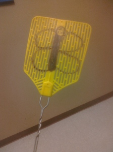
Flies didn’t evolve around flyswatters. Gotcha.Owned.


Notch and Delta in a Nutshell


Cell signaling is a very important process within multi-cellular organisms. Yet, many people grossly misunderstand how cells could communicate with one another. During the Answers in Genesis conference that stopped by Morris last year, one individual was under the impression that “cellular e-mail” must exist. “Why? Well, because it must!” *sigh* So, the pathway that I’m going to look at is Notch. Why? Well, because I must!  It’s also pretty cool, I swear.

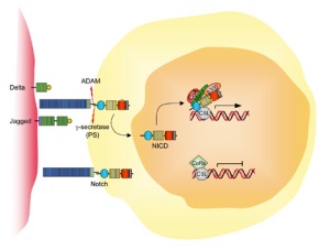
So here we have the Notch pathway. http://www.humpath.com/IMG/jpg_notch_jagged_gs_01.jpg Notch is a transmembrane protein which means it goes straight through the cell membrane and protrudes on either side. This protein acts as a form of hair-trigger. When activated it cleaves the inside part which heads off to the nucleus to play with gene expression. Notch isn’t activated by just any protein flying into it, and this is where Delta comes into play. Now the specifics vary depending on species (humans don’t actually have Delta but it’s similar and therefore Delta-like) but the purpose remains relatively the same.

**Now, I’m grossly oversimplifying the Notch pathway. I do this not because the areas I’m overlooking are not important (they are!), but this is a neurology post and it’s far too easy to get bogged down in minutia of everything and overlook the cool stuff of one area. **

So Delta and Notch touch, part of Notch breaks off and heads off into the cell to make shenanigans. What does this shenaniganary have to do with cell signaling and the nervous system? These have two functions. One is to adhere the two cells to one another. The other function is in development, in this case neuro-development. Once the cell with Delta contacts the Notch cell the piece inside breaks off and heads for the nucleus. What it does here is stop the cell from differentiating while the Delta cell remains able to do so. Notch inhibits rather than induces. It’s a bit backwards from what you would expect, but the role of Notch in neural development is to say who cannot become neural tissue.  As for the Delta cell, eventually it will move out of the epithelial layer and differentiate into a neuroblast.

If you think about it, this makes sense. If something induces neural differentiation then something should also stop it to avoid something made entirely of nervous system. Now, I can’t emphasize enough that Notch does other things. Those things just reach beyond the scope of this class. This is just a quick look at how two little things poking out of a cell can have a big impact.